|
Ethnobotanical Leaflets 13:1417-25. 2009.
Morphological and Anatomical Studies of the Leaf and Stem of Some Medicinal Plants: Stachytarpheta jamaicensis (L.) Vahl. and S. cayennensis (L.C.Rich) Schau.
1Idu, M*., 1J.O. Erhabor and 2Odia, E.A.
1Department of Botany, University of Benin, P.M.B. 1154, Benin City, Nigeria. 2Department of Botany, Ambrose Alli University, Ekpoma, Edo State, Nigeria
Issued 01 November 2009
Abstract
A comparative study of the morphological and anatomical features of the leaves and stems of Stachytarphetajamaicensis and S. cayennensis was undertaken; both species have been widely reported in several herbal medicines. The presence of angular stem and pubescent leaves in the latter distinguishes it morphologically from the former, which is characterized by smooth circular stem and glabrous leaves. The use of a light microscope revealed the presence of trichomes in the leaf of S. caynnensis but absent in S. jamaicensis. Differences in epidermal structure and stomata arrangements were also prominent features for separating between these species. Key words: Morphological, anatomical, medicinal plants, Stachytarpheta jamaicensis, Stachytarphetacayennensis, leaf, stem. Introduction The family Verbenaceae occurs mainly in the tropics and subtropics and comprises about 98 genera and 3,000 species. The Verbenaceae family may be herbs, shrubs and trees. They are economic plants and may be grown as ornamentals (Gill, 1988). Verbenaceae species are popular in traditional medicine (lyang, 2003). Stachytarpheta cayennensis is popular in traditional treatment of malaria, heartburn, ear-sores as well as eye problems such as conjunctivitis, initis and trachoma (lyang, 2003). S. jamaicensis (Brazillian tea) is used for allergies and respiratory conditions such as cold, flu, asthma, bronchitis and others, it is also used for digestive problems such as indigestion, acid reflux, ulcers, constipation, dyspepsia and (Idu et al., 2007). In Nigeria, it is known for treatment of diabetes, hypertension and bacterial infection (Ataman et al., 2006). The leaves or entire aerial parts is prepared as a hot tea for a stomach tonic to stimulate the function of the gastrointestinal tract, for dyspepsia, fever and to promote perspiration as well as for chronic liver problem (Coimbra, 1994). It is contraindicated in pregnancy and for persons with low blood pressure, as it is both abortifacient and hypotensive (Taylor, 2005). Apart from physiognomic characters, anatomical properties of plant parts are sources for taxonomic inferences in different groups of flowering plants (Edeoga et al., 2007; Guimeraes et al., 2007; Kaplan et al., 2007; Keshavarzi and Zare, 2006). The present paper is a comparative study of the morphological and anatomical characteristics of the leaves and stems of S. jamaicensis and S. cayennensis.
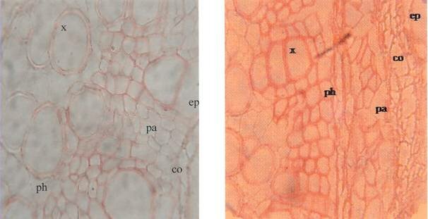
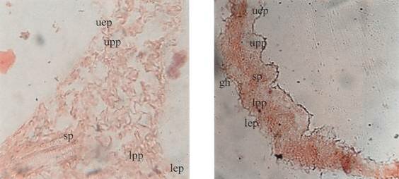
Materials and MethodsSamples of S. cayennensis and S. jamaicensis were collected from a home garden in BDPA, Ugbowo, Benin City, Edo State, Nigeria. They were properly identified using literature (Akobundu and Agyakwa, 1998). Morphological assessment was by physical observation and measurement of physiognomic features of their fresh leaf and stem specimens. For the anatomical studies, the fresh samples were fixed in Booing fixative and cross sections obtained using a microtome (Johansen, 1940). The sections were independently stained with heamatoxyline and safranin. A light microscope was used to view the slides and adjusted to finest resolution (x40). Microphotographs were obtained using a Nikkon digital camera focused through the microscope eyepiece. Results and DiscussionMorphologically, S. jamaicensis is an erect, straggling perennial herb, 0.6-0.9 m, stem smooth and woody. Leaves glabrous, 10.0 cm x 4.8 cm ovate or oblong to elliptic. While S. cayennensis is an erect perennial herb, branched and woody at the base. Stem is 4-angled, 1.1-1.3 m with free branching stems covered with short hairs. Leaves 8.0 x 3.8 cm ovate toelliptic and pubescent. Leaf sections of both species (Fig. 1) revealed similar structural pattern or arrangement. The epidermis is covered with thick cuticle, palisade parenchyma is found on both sides of the spongy parenchyma. Stomata are found on both sides of the leaf (amphistomatic). Their stem sections (Fig. 2) showed an outer thick cuticle layer followed by a single layered epidermis and chlorochymatous tissues covering a small area under the epidermis. The collenchyma cells are thick walled and located between epidermis and chlorochymatic layers. There is evidence of secondary growth and vascular bundles are scattered in a circular form with phloem on the outer and xylem on inner sides. Vascular bundles are surrounded with a single layer of parenchyma cell. The pith is parenchymatous. However, the major differences in anatomical features in the stems and leaves of both species are outlined in Table 1. Table 1: Comparison of Anatomical Characteristics between S. jamaicensis and S. cayennensis leaf and stem.
A B Fig. 1. Photomicrographs of T.S. of stem (x40). A & B, S. jamaicensis and S. cayennensis respectively showing structural pattern. Key: ep- Epidermis, co- Collenchyma, pa- Parenchyma, ph- Phloem, x- Xylem.
A B Fig. 2. Photomicrographs of sections of leaf (x40). A & B, Transverse section of S. jamaicensis and S. cayennensis respectively showing the structural arrangement of layers;
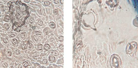
C D C & D, Surface section (Abaxial) of S. jamaicensis and S. cayennensiss respectively showing density of stomata and presence of trichomes. Key: uep- Upper epidermis, upp- Upper palisade parenchyma, sp- Spongy parenchyma, lpp- Lower palisade parenchyma, lep- Lower epidermis, gh- Glandular hair, st- Stomata, ep- epidermis.
The morphological features observed in both specimens used in the present study were consistent with the descriptions reported by Akobundu and Agyakwa (1998). The major distinguishing features include the presence of angular stem in S. cayennensis while that of S. jamaicensis is rather circular. Also, the leaf and stem of the former are pubescent, but glabrous in the latter. Transverse sections of the stems (Fig. 1) revealed the structural arrangements in both species are very similar; an outer epidermal layer followed by 2-5 layers of collenchymatous cells. The vascular bundles are oval and concentric, with the xylem towards the center surrounded by the phloem. The evidence of secondary growth in the stem sections is congruent with the woody morphological description earlier reported by Akobundu and Agyakwa (1998). Observable differences were more pronounced in sections of their leaves (Fig. 2). The upper epidermis of S. jamaicensis leaf was thicker compared to that of S. cayennensis while stomata were more abundant in the abaxial surface of S. cayennensis (Table 1). The most outstanding anatomical feature separating between the species is the presence of trichomes in the surface section (abaxial) of S. cayennensis leaf but distinctly lacking in S. jamaicensis.
ReferencesAkobundu, I.O and Agyakwa, C.N. 1998. A Hand Book on West African Weeds. International Institute of Tropical Agriculture Publication Ibadan, Nigeria, 564p. Ataman, J.E., Idu, . Odia, A.E., Omogbai, E.K.I., Amaechina, F. Akhigbe, A.O. and Ebite, L.E. 2006. Histopathological effects of Stachytarpheta cayennensis (L.) Vahl. on Wister rats. Pakistan Journal of Biological Sciences, 9: 477-482. Coimbra, R. 1994. Manual de Fitoterapia. 2nd Edition, Editoria cejup Belem, Brazil 4p. Edeoga, H.O., Omosun, G., Osuagwu, G.G.E. and Emezue, O.O. 2007. Microscopic anatomy and histochemistry of stem and root of some Mimosa species. (Leguminosae- Mimosoideae). Asian Journal of Plant Sciences, 6(4): 688-691. Gill, L.S. 1988. Taxonomy of Flowering Plants. Africana-FEP Publishers Limited, Bamenda, Cameroon. 388p. Guimeraes, A.C., Kuster, R.M., Amaral, A.F., Ferreira, J.P. and Siani, A.C. 2007. Histological study of the leaf and stem of the Amazonian medicinal Mistletoe Cladocolea micrantha (Loranthaceae). International Journal of Botany, 3(2): 218-221. Idu, M., Omogbai, E.K.I., Aghimien, G.E., Amaechina, F., Timothy, O. and. Omonigho, S.E. 2007. Preliminary phytochemistry and antimicrobial properties of Stachytarpheta jamaicensis (Linn.) Vahl. stem. Research Journal of Medicinal Plants, 1(4): 145-153. Iyang, E. 2003. Ethnobotany. Conventional and Traditional Uses of Plants. The Verdict Press, Uyo, Akwa-Ibom State, Nigeria. 191p. Johansen, D.A. 1940. Plant Micro-technique. McGraw- Hill Publishers, New-York. 523p. Kaplan, A., Hasanoglu, A. and Ince, I.A. 2007. Morphological, anatomical and palynological properties of some Turkish Veronica species (Scrophulariaceae). International Journal of Botany, 3(1): 23-32. Keshavarzi, M. and Zare, G. 2006. Anatomcial study of Salicornieae Dumort. (Chenopodiaceae Vent.) Native to Iran. International Journal of Botany, 2(3): 278-285. Taylor, S. (2005). The Healing Power of Rainforest Herbs. No Square Area Publishers, Garden City, New York, 519p.
|